Howell jolly body
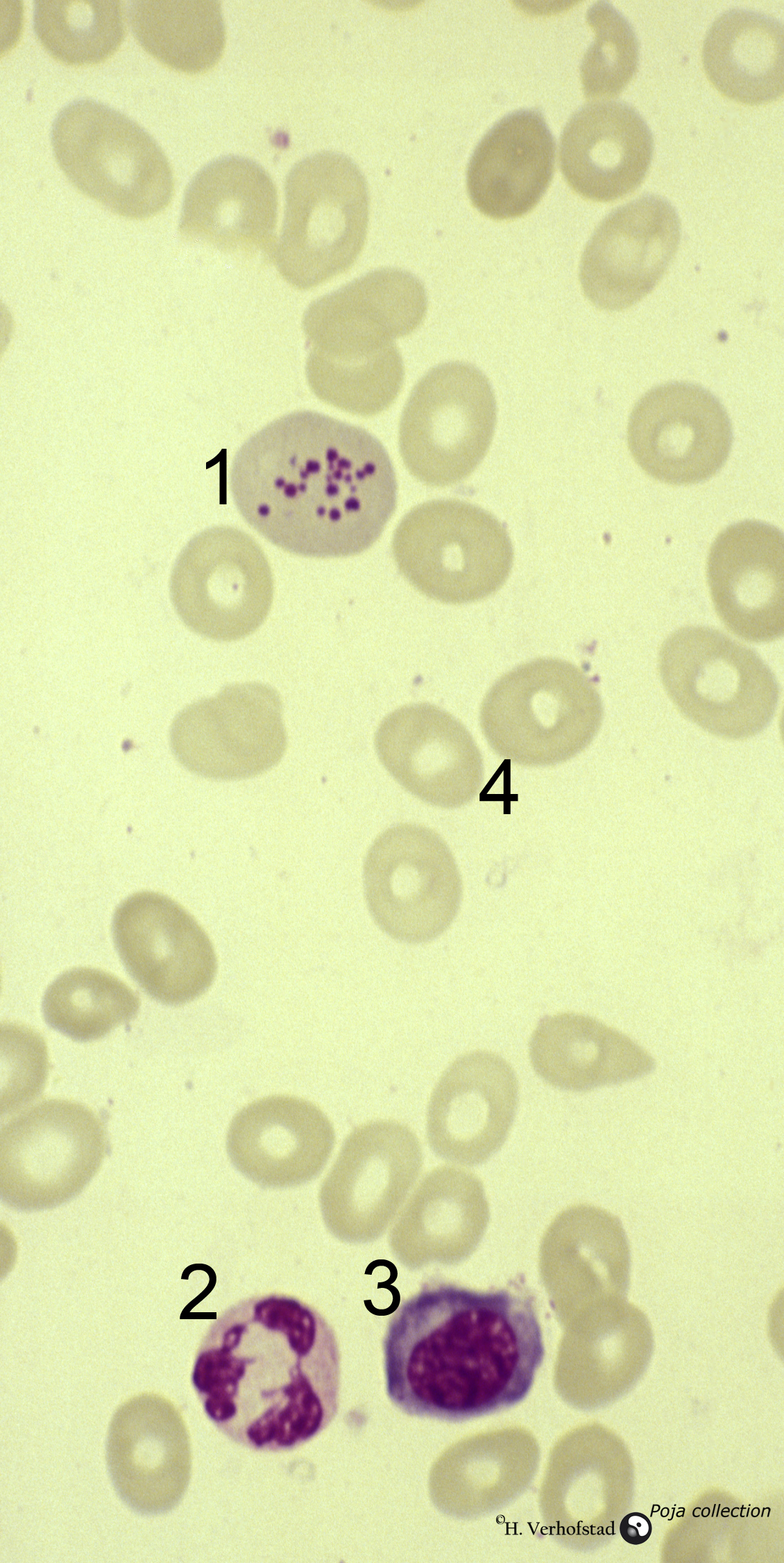
With HIV infections, the presence of detached nuclear fragments in the cytoplasm of neutrophils that bear a resemblance to the nuclear remnants of red cells known as Howell-Jolly bodies can be found. These unusual inclusions should be differentiated from other intracytoplasmic inclusions, such as those that may be seen in infections or in rare.

Basophilic stippling and HowellJolly bodies
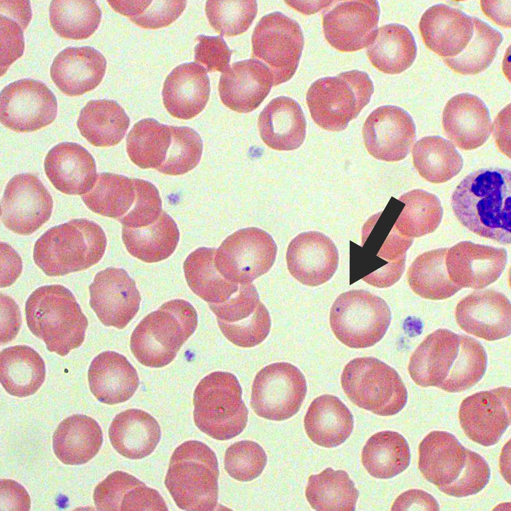
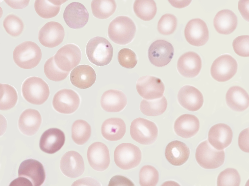
Under Wright/Romanowksy stains, Howell-Jolly Bodies appear as dark blue/purple round inclusions located at the periphery of the RBC. They usually present as a single inclusion inside the cell. Howell-Jolly Bodies are also visible under supravital stains. 1-4. Inclusion composition: 2,3. Nuclear fragments/remnants made up of DNA 1-4

HowellJolly bodies in the peripheral blood smear (case 3) Download Scientific Diagram
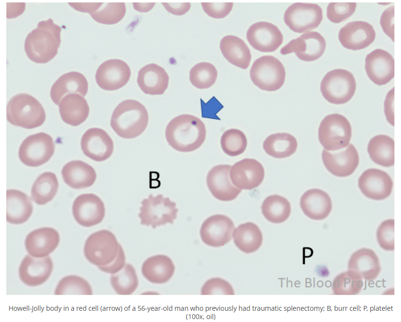
Howell-Jolly bodies During maturation in bone marrow, erythrocytes normally expel their nuclei but sometimes a small portion of DNA remains In healthy people, Howell-Jolly bodies are pitted out by spleen during erythrocyte circulation Etiology Howell-Jolly bodies persist in those with functional hyposplenia or asplenia:
HowellJolly bodies in reticulocytes in peripheral blood smear (human) Eccles Health Sciences
Howell-Jolly bodies occur where there is no spleen or an non-functioning spleen, referred to as asplenia. They are usually one of these at most in a red cell, round, dark purple to red in color and often located peripherally on the red blood cell. If a patient comes in with sepsis, fever, headache and myalgias, meningitis is very likely.

HowellJolly Bodies Cells and Smears
Howell-Jolly bodies are nuclear remnants found in red blood cells (erythrocytes) under various pathological states. They most commonly present in patients with absent or impaired function of the spleen; this is because one of the spleen's functions is to filter deranged blood cells and remove the in.
Howell Jolly Bodies Test Findings MedSchool
Instant-Address, Phone, Age & More A Jolly- Search Now. Get A Jolly's Phone, Email, Social Profiles - No Hit No Fee!
HowellJolly body
Objectives: Identify the etiology of functional asplenism. Describe the presentation of a patient with functional asplenism. Review the treatment and management options available for functional asplenism.
What is a HowellJolly body? • The Blood Project
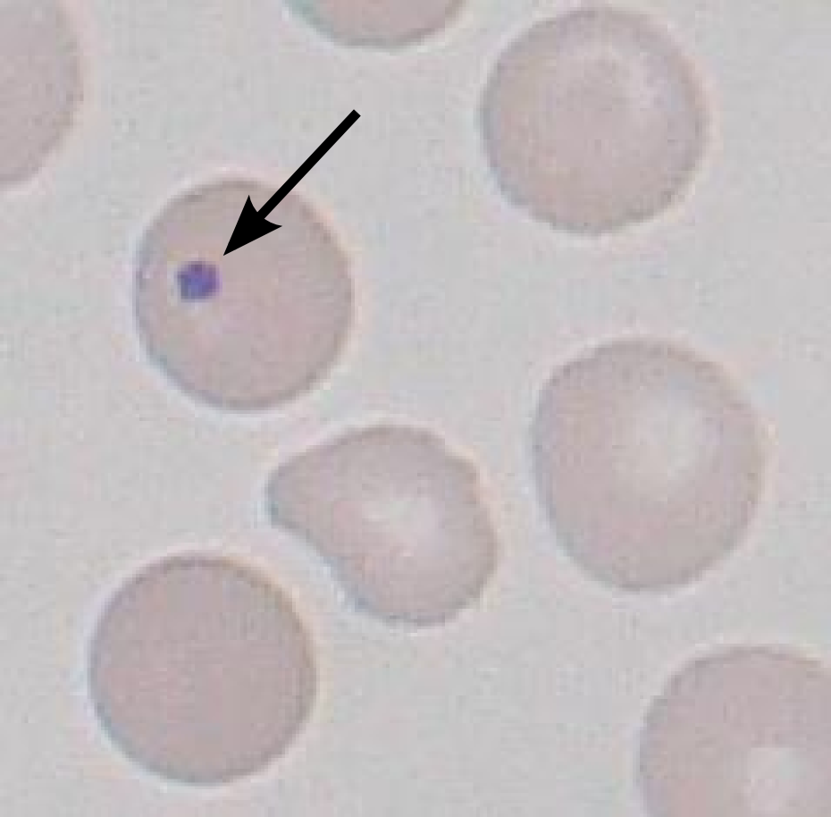
Howell-Jolly bodies are small round purple inclusions in RBCs about 1 μm in diameter. Compared to Pappenheimer bodies, Howell-Jolly bodies are larger in size, have smooth outlines, typically one per RBC, and are comprised of DNA. Platelet overlying a red cell, Pappenheimer bodies, artifact. They are formed in the process of red cell nuclear.
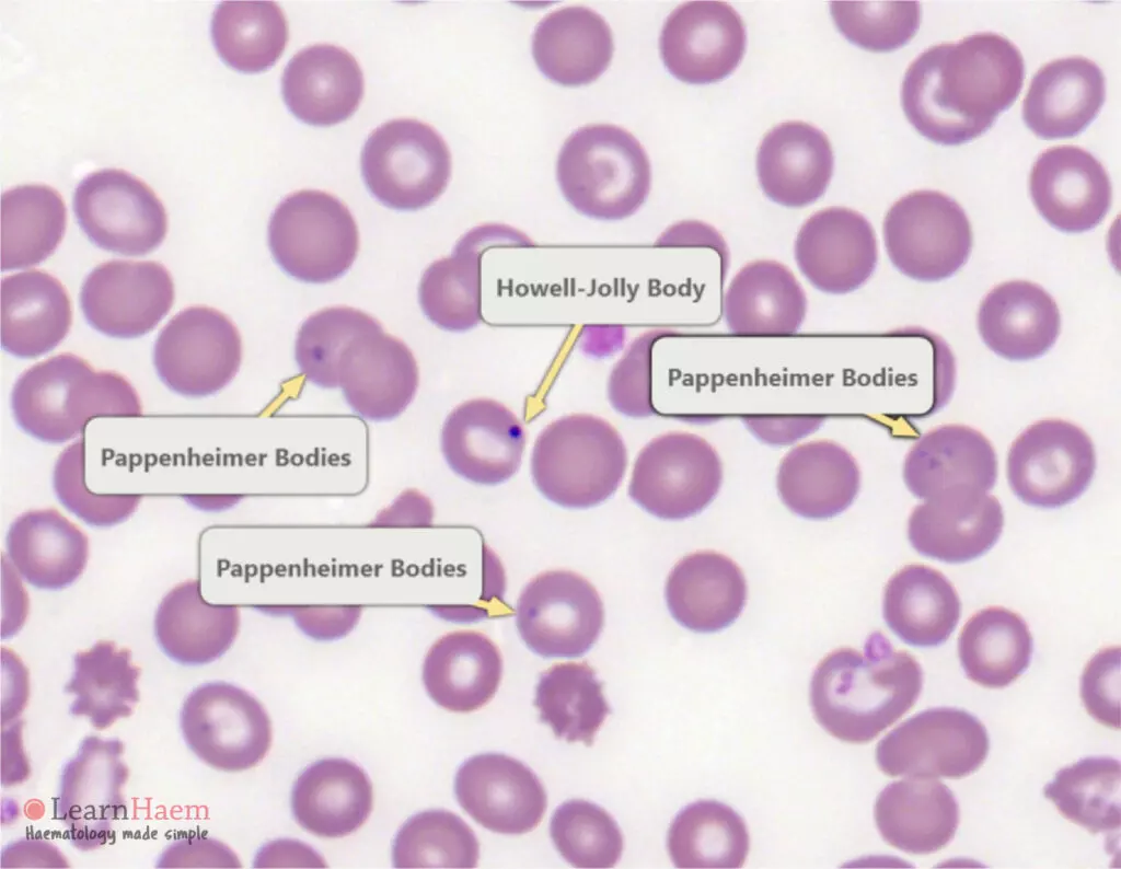
Pappenheimer Bodies Vs Basophilic Stippling
Howell-Jolly bodies are small (0.5-1 micron) purple inclusions that contain DNA. They are thought to represent chromosomes that have separated from the mitotic spindle that are left behind when the red cell nucleus is extruded. These inclusions are generally removed by the spleen.
Howell Jolly bodies
Howell-Jolly bodies are DNA-containing inclusions found after erythrocyte maturation. The composition of the DNA is still unknown to this day. However, studies show that they are of centromeric origin.

Howell Jolly Body found tonight
Home Hematology Cell morphology Red blood cells Inclusions Inclusions Red blood cell inclusions can arise from a variety of sources. Correct identification of these abnormalities is important since it can provide insights into metabolic, physiologic, and pathologic conditions affecting the red blood cells. Basophilic stippling

HowellJolly bodies in neutrophils Blood Academy
David S Rosenthal, MD Section Editor: Robert A Brodsky, MD Deputy Editor: Jennifer S Tirnauer, MD Literature review current through: Nov 2023. This topic last updated: Oct 05, 2023. INTRODUCTION Examination of the peripheral blood smear is an inexpensive but powerful diagnostic tool in both children and adults.

Why do you see HowellJolly bodies in sickle cell anemia? Pathology Student
Howell-Jolly Body . On these Wright-stained peripheral blood smears the small dark spheres within red cells are Howell-Jolly bodies. Note the variation. in size. These bodies represent residual nuclear DNA which, under normal circumstances, is removed by the spleen. There can . be more than one H-J body within a red cell.
HowellJollylike bodies in neutrophils
The inclusions persisted at stable frequency after treatment. Howell-Jolly-like bodies in granulocytes arise secondary to stressed granulopoiesis induced by immunosuppressive drugs, viral infection, or chemotherapy, and must be differentiated from other neutrophil inclusions such as those observed in intracellular bacterial infections.
Histology, Howell Jolly Bodies Article StatPearls
Prime Try Before You Buy is now available for eligible Prime members! Explore men's & women's new arrivals, shop latest sales & deals, and everyday essentials
Sickle Cell Disease/Sickle Cell Anemia Stepwards
Howell-Jolly bodies are nuclear remnants. They are small, round cytoplasmic inclusions that stain purple on a Romanowsky stain. They are regularly present after splenectomy and when there is splenic atrophy ( Fig. 5-57 ). They may be seen in a small percentage of red cells in pernicious anaemia.
